SCIM_Tif
Store raw data from ScanImage Tif images
Usage
OBJ = SCIM_Tif(FILENAME, CHS, CAL)
Arguments
FILENAMEis the original filename (including the path) of thisSCIM_Tifobject.CHSis a scalar structure that contains information about the meaning of the image channels.CALis a scalarCalibrationPixelSizeobject.
Details
SCIM_Tif objects are used to contain all raw image data and metadata from ScanImage Tif images. This class relies on and includes existing code provided with the ScanImage microscope software. For more information on ScanImage, please visit http://www.scanimage.org, or refer to Pologruto et al. (2010), BioMed Eng OnLine 2(1):1-9.
See Also
RawImgclass documentationMetadataclass documentationMetadataquick start guideCalibrationPixelSizeclass documentationCalibrationPixelSizequick start guide
Examples
The following examples require the sample images and other files, which can be downloaded manually, from the University of Zurich website (http://www.pharma.uzh.ch/en/research/functionalimaging/CHIPS.html), or automatically, by running the function utils.download_example_imgs().
Create a SCIM_Tif object interactively
The following example will illustrate the process of creating a SCIM_Tif object interactively. It is possible to do so by using the from_files() method, rather than the constructor (as is demonstrated in this section).
% Call the SCIM_Tif constructor
st001 = SCIM_Tif()
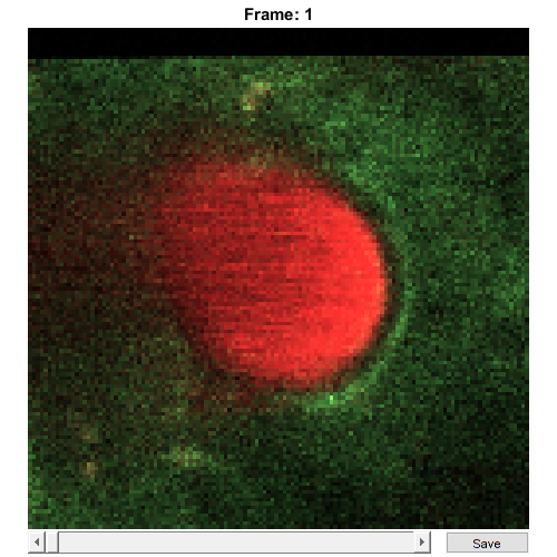
Use the interactive dialogue box to select the raw image file xsectscan_scim.tif, which should be located in the subfolder tests>res, within the CHIPS root directory:.

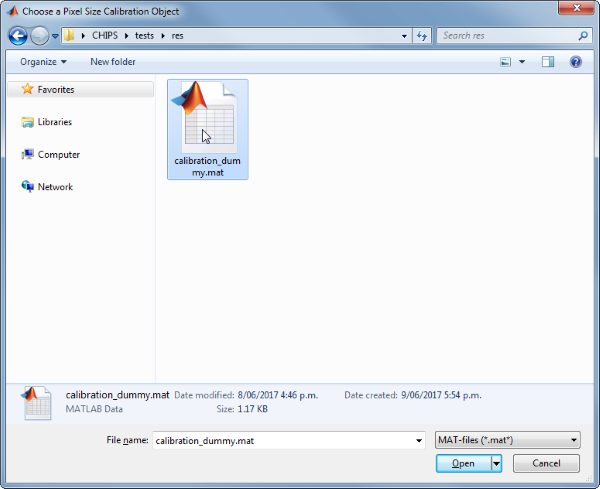
Use the interactive dialogue box to select the dummy calibration (calibration_dummy.mat):
The next stage is to define the ‘meaning’ of the image channels. The first channel represents a cellular sensor, and the second channel represents the blood plasma. Press six and enter, followed by one and then enter, to complete the selection.
----- What is shown on channel 1? -----
  >> 0) <blank>
     1) blood_plasma
     2) blood_rbcs
     3) Ca_Cyto_Astro
     4) Ca_Memb_Astro
     5) Ca_Neuron
     6) cellular_signal
     7) FRET_ratio
Answer: 6
----- What is shown on channel 2? -----
  >> 0) <blank>
     1) blood_plasma
     2) blood_rbcs
     3) Ca_Cyto_Astro
     4) Ca_Memb_Astro
     5) Ca_Neuron
     6) cellular_signal
     7) FRET_ratio
Answer: 1
We have now created a SCIM_Tif object interactively.
st001 =
SCIM_Tif with properties:
            filename: 'C:\...'
          isDenoised: 0
   isMotionCorrected: 0
   metadata_original: [1x1 struct]
                name: 'xsectscan_scim'
             rawdata: [4-D uint16]
                  t0: 0
            metadata: [1x1 Metadata]
The process is almost exactly the same to create an array of SCIM_Tif objects; when the software prompts you to select one or more raw images, simply select multiple images by using either the shift or control key.
Create a SCIM_Tif object without any interaction
% Specify the full path to the raw image object
fnST002 = fullfile(utils.CHIPS_rootdir, 'tests', 'res', ...
    'xsectscan_scim.tif');

% Specify the channels relevant for this raw image
channels = struct('cellular_signal', 1, 'blood_plasma', 2);

% Load the CalibrationPixelSize object
fnCalibration = fullfile(utils.CHIPS_rootdir, 'tests', 'res', ...
    'calibration_dummy.mat');
calibration = CalibrationPixelSize.load(fnCalibration);

% Create the SCIM_Tif object without any interaction
st002 = SCIM_Tif(fnST002, channels, calibration);

% View the SCIM_Tif object
st002.plot()
Opening xsectscan_scim.tif: 100% [=================================]